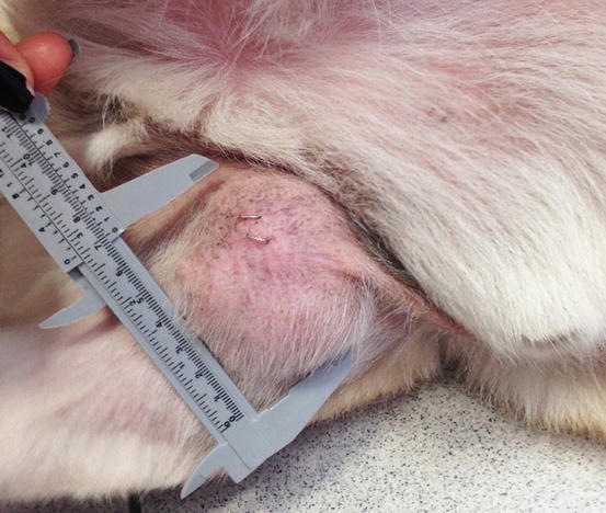

Think about how big a cat’s head is, and you’ll see that is a big tumor. I know this is a blog about dog cancer, but these cases illustrate exactly why it can be problematic to wait too long to take action – no matter what species we’re talking about.Įarlier in the week, I met Tiger, a big 16 year old cat with a recurrent tumor on his lower jaw, up front by the incisors. This week I saw two cases that really depressed and frustrated me. I feel we are waiting too long, for too many masses.
#Spindle cell sarcoma in dogs skin#
While many skin masses are benign, some of these masses may also be slow-growing malignant tumors and it is better to remove them early, when it is more likely that the tumor can be completely removed with wide margins with the FIRST surgery.
#Spindle cell sarcoma in dogs plus#
In my veterinary training, I was taught that if a mass is not growing or changing in appearance, it is likely ok to do nothing and leave it – “just continue to monitor.” It’s been ten plus years since I started my medical oncology residency, and from my experience treating dogs and cats with cancer, that is not always the best advice. But how long is ok? What size is too big? Are there actual guidelines? I too have advised that to my pet Guardians. The relationship between spindle cell tumors of the anterior uvea of dogs, altered neural crest, blue iris color, and ultraviolet radiation has not yet been fully elucidated.Have you been told to “just watch” a lump or mass on your dog by a veterinarian? I wouldn’t be surprised if you have. These tumors are morphologically and immunohistochemically most consistent with schwannoma. Electron microscopy revealed intermittent basal laminae between cells. Tumors were variably positive for PGP 9.5, laminin, gadd45, p53, PCNA, anti-UVssDNA, and TERT. All tumors were negative for SMA, desmin, Melan A, and MITF-1. Nine of 13 tumors exhibited GFAP immunopositivity. All tumors were positive when immunostained for vimentin and S-100. All tumors occurred in the iris with or without ciliary body involvement and were composed of spindle cells arranged in fascicles and whorls (variable Antoni A and B behavior). Immunohistochemical staining included alpha-smooth muscle actin (SMA), vimentin, S-100, desmin, glial fibrillary acidic protein (GFAP), Melan A, microphthalmic transcription factor (MITF-1), protein gene product 9.5 (PGP 9.5), laminin, gadd45, p53, proliferating cell nuclear antigen (PCNA), anti-UVssDNA (antibody for detection of (6-4)-dipyrimidine photoproducts), and telomerase reverse transcriptase (TERT). Light microscopic evaluation (all dogs) and electron microscopy (2 dogs) were performed. Siberian Husky and Siberian Husky mix dogs were overrepresented (10/13 dogs, overall median age 10 years). Thirteen tumors were identified from the 4,007 canine ocular samples examined at the Comparative Ocular Pathology Laboratory of Wisconsin between 19. Immunohistochemical techniques were used to investigate the origin of a spindle cell tumor in the anterior uveal tract of dogs and the influence of ultraviolet radiation on the development of this tumor.


 0 kommentar(er)
